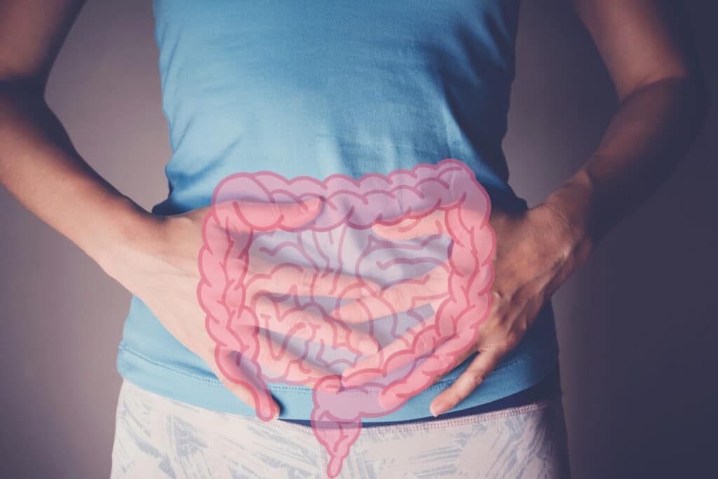
|
|
Journal of Spirochetal and Tick-borne
DiseasesVolume 5, Spring/Summer 1998
An Understanding of Laboratory Testing for
Lyme Disease
INTRODUCTION
While Lyme disease is
a clinical diagnosis, the laboratory can provide useful and necessary information
for the diagnostic process. The question is how does one use the laboratory
in the most appropriate and cost effective manner? The following examples
stress the need for an understanding of the tests available and when and why
to use them.
HYPOTHETICAL CASE REPORT
A female patient in New
Jersey presents to a university rheumatologist with symptoms of arthralgia
and myalgia, fatigue and malaise, rash, photosensitivity, mild cognitive dysfunction,
and nonspecific gastrointestinal complaints. After a thorough physical examination,
the rheumatologist orders a WBC, multiple analyte chemistry panel, sedimentation
rate, ANA with a reflux to ENA if the ANA is positive, rheumatoid factor,
anticardiolipin, C3, C4, VDRL, urinalysis, and perhaps some joint x-rays if
the physical diagnosis is supportive.
This same patient also
noted that one of her neighbors contracted Lyme disease and believes she may
have it as well. The rheumatologist then added a screening test for Lyme disease,
either ELISA or an IFA quantitative (titer) test. If the ELISA or IFA were
negative, the chance of a diagnosis of Lyme disease in this patient would
be remote because the current dogma (1-3) is that Lyme
disease is a rather rare event in most parts of the country, especially in
the absence of a positive ELISA or IFA screening test.
BACKGROUND
There have been some
good reviews (4-6) prior to 1994 on the laboratory aspects
of diagnosis, but most of these were written before the politicizing of the
diagnostic process during the CDC/ASPHLD meeting in Dearborn Michigan (7).
Prior to 1994, the CDC recognized Lyme disease from a set of clinical symptoms
and a general set of laboratory findings. A certain combination of these criteria
would lead to diagnosis of Lyme disease that could be reported to the CDC.
The Dearborn meeting changed that.
The original clinical
case definition (8) from the CDC for Public Health Surveillance
and reporting of Lyme disease was:
Clinical Criteria:
- Erythema Migrans;
or
- At least one late
manifestation of musculoskeletal, nervous or cardiovascular system disorder;
and laboratory confirmation.
Laboratory Criteria:
- Isolation of Borrelia
burgdorferi from clinical specimens; or
- Demonstration of diagnostic
levels of IgM and IgG antibodies to the spirochete in serum or CSF (Western
blot, ELISA, IFA), or
- Significant changes
in IgM or IgG antibody response to Borrelia burgdorferi in paired
acute- and convalescent-phase serum samples.
These criteria placed
great emphasis on the presence of an Erythema Migrans (EM) rash. It is usually
accepted that a physician's diagnosis of an EM on a patient from an endemic
area is extremely useful for diagnosing Lyme disease; almost a third of the
patients actually do not have an EM (9-11). In addition,
the variability of the EM rash (12,13),
such as its duration, nonpruritic and nonpainful nature, and its location
in obscure areas (axilla and hair regions) inhibit its use as a consistent
diagnostic marker.
In 1995, the CDC added
the additional recommendation from the CDC/ASPHLD meeting (7)
of a two-tiered approach for reporting active disease and previous infection.
That requirement means that a positive sensitive ELISA/IFA must be
followed by a positive Western blot with a defined number of approved antibody
bands. If the intention were only for public health surveillance and reporting
of disease, these changes would not have caused a problem. Unfortunately,
these recommendations became the standard in most areas and especially with
insurance companies. That was unfortunate because the Dearborn meeting was
not supposed to be about setting national standards for Lyme disease diagnosis;
rather, it was to be a discussion regarding the Western blot during early
Lyme disease. The majority of patient samples used to set the criteria were
from patients being followed for four months following their diagnoses. The
patients considered for entry in the study had an EM rash and either arthritis
or neuroborreliosis.
Lyme disease is a problematic
diagnosis because it is a complicated clinical entity. The position by the
CDC makes it more complex. Some patients do not elicit an antibody response
great enough to be positive by the currently available ELISA assays. Recent
studies (14) by the group responsible for Lyme disease
proficiency testing for the College of American Pathologists (CAP) came to
the conclusion that the currently available ELISA assays for Lyme disease
do not have adequate sensitivity to meet the two-tiered approach recommended
by the CDC/ASPHLD group (7). In addition, Bakken et al
(14) stated that a screening test must have sensitivity
>95% to adequately screen for Lyme disease and that the currently available
ELISA tests do not meet this criteria. Furthermore, if patients are treated
early with antibiotics, their antibody response may be reduced or curtailed
(15). The initial mild flu-like symptoms may be overlooked.
Later, if the symptoms return, most of the antibody markers have disappeared.
The picture is not entirely bleak if Lyme disease is approached for what it
is: a complicated clinical entity, which requires multiple laboratory
tests to assist in the diagnosis. Thus, if clinicians use multiple tests
(ie, both screening and confirmatory Western blot assays, antigen-capture
and PCR), as they do in other disease entities, there will be fewer problems
with the diagnosis and fewer patients will be missed.
The presence of detectable
spirochetes in infected tissue is rare. The characteristic sparsity of organisms
contributes to the difficulty of getting blood or tissue to grow the Lyme
bacterium (15). A positive culture may not be a predictor
of an antibody response. Rawlings (16) followed a group
of 14 patients in which she was able to culture B. burgdorferi, but
only 3 of those patients had positive antibody titers. Aguero-Rosenfeld et
al (12,13) showed that only 70%
of the documented Lyme patients in their study had a significant antibody
response. They suggested that the degree of antibody response might be related
to the length of time the EM rash persists. They also saw only a 64% rate
of IgM to IgG seroconversion.
Early reports suggested
that considerable interlaboratory and intralaboratory variability exist in
Lyme disease testing (17-19). However, a review of the
1996 Lyme proficiency results by CAP (College of American Pathologists) and
those by New York State demonstrates comparable agreement between the laboratories,
similar to other bacterial infections and autoimmune conditions.
Table 1. Assays
for Lyme disease
Direct
Biopsy
Culture
Antibody Assays
IFA
ELISA
Western blot
Antigen Assays
Antigen-Capture
PCR
RESULTS AND DISCUSSION
Table 1 presents the
types of tests that are most commonly available for Lyme disease. To provide
adequate support for the clinical evaluation, multiple tests should be used.
Not only is a correct diagnosis advantageous for the patient, but also ultimately
is the most cost effective.
Indirect fluorescent
antibody (IFA)
B. burgdorferi
spirochetes are affixed to glass slides and usually a fluorescent-conjugated
goat antihuman immunoglobulin of either IgM or IgG specificity is used (20).
Tests for Lyme disease using IFA have received mixed reviews and some authors
believe that the interpretations of IFA assays are overly subjective and that
the tests are either functionally insensitive for Lyme-specific antibodies
or display considerable cross-reactions with antibodies to other spirochetal
organisms (21,22). Magnarelli et
al (23,24) and Mitchell et al (20)
supported IFA if used in conjunction with a clinical evaluation. Mitchell's
study with the IgM IFA showed excellent specificity and no observed cross-reactivity
with infectious mononucleosis (n = 20), rheumatoid arthritis (n = 19), systemic
lupus (n = 22), syphilis (n = 13), streptococcal sequelae (n = 20) or healthy
subjects. Mitchell related the success of this test to the quality of the
substrate slides and the level of experience of the technologists, and concludes
that IFA microscopy becomes less subjective with experience.
Enzyme-linked immunosorbant
assay
ELISA for B. burgdorferi
has been available since 1984 (25). Most commercial assays
use a whole cell sonicate of B. burgdorferi. Complete descriptions
of methods for a Lyme ELISA can be found in the publications by Craft et al
(25), Magnarelli et al (23), and
Russell et al (21). Standard ELISA techniques have been
employed (26) in all these assays.
There are a large number
of commercial ELISA tests available. A review of past proficiency events by
CAP and the NYS Health Department show the relationship between the various
tests. Most commercial ELISA tests have comparable sensitivity and specificity
because they were made to compare to one another for the FDA 510K process.
However, most are inadequate as a screening test because they were not designed
by the manufacturers to be sensitive at the 95% level, which is required for
screening (14). A substantial change in the 510K approval
process would be required to make the ELISAs for Lyme disease diagnosis more
sensitive.
The goal for a new generation
of ELISAs should be sensitivity for the more unique and specific B. burgdorferi
antigens that are visualized in the Western blot (Figure 1). They are Osp
A (31 kDa), Osp B (34 kDa), Osp C (23-25 kDa), 39 kDa, and 93 kDa (27-32).
Initially, some investigators identified 93 kDa as 94 kDa and Osp C as 22
kDa. While most ELISAs do have reactivity to these antigens, because they
are prepared with a sonicate of B. burgdorferi, they also have reactivity
against 41 kDa, 58 kDa, 66 kDa, and 73 kDa. While the later antigens are components
of B. burgdorferi, they also have considerable cross-reactivity to
other spirochetes, heat-shock proteins, and some viruses (33).
All borderline and positive
ELISA assays (polyvalent, IgG only, and IgM only) for Lyme disease must be
confirmed by a high quality Western blot for B. burgdorferi. A 56%
false-negative rate, depending upon the commercial kit, was found by Luger
and Krause (18), as compared to their own clinical diagnoses.
Golightly et al (34) saw a lack of sensitivity with a
70% false-negative rate in early Lyme disease and from 4% to 46% with late
manifestations of Lyme disease. These results support the necessity of Western
blot confirmation for both positive and negative Lyme ELISA.
B. burgdorferi
Western blotting
Figure 1.
Western blots of B. burgdorferi B31 strain: The blots were:
(1) stained with amino black, (2) reacted with rabbit antisera, (3) with
goat antisera, (4-7) with various monoclonal antibodies, and (8) with pooled
patient sera. Reproduced by permission from Ma B, Christen B, Leung D, Vigo-Pelfrey
C. Serodiagnosis of Lyme borreliosis by Western immunoblot: reactivity of
various significant antibodies against B. burgdorferi. J Clin Microbiol
1992; 30: 370-76.
The immunoblot or Western
blot (Figure 1) for B. burgdorferi is the most useful antibody test
available when performed in a quality laboratory by experienced testing personnel.
It is necessary to evaluate separately both the IgM and IgG antibodies of
B. burgdorferi. The study by Ma et al (35) gives
an excellent overview of the technique and provides comprehensive information
about the antibodies seen in Lyme disease patients versus the normal and non-Lyme
disease groups.
Figure 2.
Western blots to B. burgdorferi from various patients with
Lyme disease Lanes 1-48 are IgG/IgM blots from clinically confirmed patients
with various levels of antibodies. Lanes 48-57 are IgM-only blots. Reproduced
by permission from Ma B, Christen B, Leung D, Vigo-Pelfrey C. Serodiagnosis
of Lyme borreliosis by Western immunoblot reactivity of various significant
antibodies against B. burgdorferi. J Clin Microbial 1992; 30: 370-376.
Figure 2 illustrates
a group of IgG-IgM Western blots (lanes 1-48) from clinically confirmed Lyme
patients with various levels of antibodies to B. burgdorferi. In
this figure are IgM Western blots to B. burgdorferi (lanes 48-57).
While some of these patients have different patterns of antibody reactivity,
all were confirmed, clinically positive Lyme patients with physician-diagnosed
EM. The variability in the Western blot is characteristic of the variability
observed in the immune response of other diseases (eg, Hashimoto's thyroiditis,
SLE, Sjogren's syndrome, scleroderma). Our own clinical study of 186 defined
patients and 320 negative controls (Figure 3) demonstrated excellent sensitivity
and specificity for IgM using any two of the following bands: 23-25 kDa (Osp
C), 31 kDa (Osp A), 34 kDa (Osp B), 39 kDa and 41 kDa (35).
This study also demonstrated good specificity and sensitivity for IgG using
any two of the above bands. The 83/93 kDa antibody could also be included
as one of two IgG bands.
Figure 3.
Comparison of antibody reactivity to various B. burgdorferi antigens.
The dark bars are from 186 patients with clinically confirmed Lyme disease
and the light bars are from 320 normal controls. Reproduced by permission
from Ma B, Christen B, Leung D, Vigo-Pelfrey C. Serodiagnosis of Lyme borreliosis
by Western immunoblot: reactivity of various significant antibodies against
B. burgdorferi. J Clin Microbial 1992; 30: 370-76.
It is difficult for each
laboratory to perform clinical studies and establish its own ranges for normal
and disease populations. For this reason, the CDC assembled a group of academic
scientists with the assistance of the FDA and the Association of State and
Territorial Public Health Laboratory Directors (ASTPHLD) to reach a consensus
on certain criteria for the Western blot. After several meetings they arrived
at the CDC/ASPHLD consensus criteria presented in Table 2 (7,36).
These criteria were based in large part on the work of Dressler et al (37),
using well-defined patients with active Lyme arthritis or neuroborreliosis.
Interestingly, in their publication none of the three CDC/ASPHLD recommended
strains of B. burgdorferi (B31, 297 and 2591) were used. Rather,
they used G39/40 with a 10% acrylamide gel, although a gel with less than
11% of acrylamide does not have enough resolution nor definition of all the
important antigens of B. burgdorferi.
The criteria for a positive
Western blot to B. burgdorferi developed by the CDC/ASPHLD are very
conservative and require 5 of 10 antibody bands for IgG positivity; the original
recommendations do not even recognize equivocal or borderline results if less
than five bands are detected. Their cut-off assumes that all Lyme patients
have similar immune systems. They ignore the diversity of the immune response
seen in other diseases. Their studies were problematic in that they primarily
focused on patients with early (usually within four months of an EM) Lyme
disease. They drew blood in most patients every two weeks during this four-month
period and any positive event (five out of ten bands) was counted as a positive
patient, even if they were negative at a different time of the study. In addition,
the criteria include antibodies to 41 kDa, a common antigen of most flagella-bearing
organisms, and exclude two of the most important and specific antigens, 31
kDa (Osp A) and 34 kDa (Osp B), which appear later in the response. A review
by Hilton et al (38) in a group of 50 patients with confirmed
Lyme disease showed that they would have missed 4 patients by excluding 31
kDa (Osp A) and 34 kDa (Osp B). The author's own laboratory would have missed
2 of 18 proficiency samples by excluding antibodies to these two antigens.
Engstrom et al (11)
and Aguero-Rosenfeld et al (12,13)
confirmed that almost one-third of all Lyme patients are IgG seronegative
during the first year. Two years after physician-diagnosed EMs, 45% of the
patients were negative by ELISA. In another study, Aguero-Rosenfeld et al
(13) showed that the ELISA response declined much more
rapidly than the Western blot response. Their study also demonstrated that
the two-step protocol of the CDC/ASPHLD criteria would fail to confirm infection
in some patients with culture-proven EM. Furthermore, although a majority
(89%) of patients with EM rash developed IgG antibodies detected by Western
blot sometime during disease, only 22% were positive by the criteria of the
CDC/ASPHLD (13). The Engstrom et al study (11)
did not use the IgG blot criteria of the CDC/ASPHLD. They found that 2 of
5 bands gave them a specificity of 93 to 96% and a sensitivity of 100% in
the 70% of patients who made antibody. This might imply that they would have
had even less sensitivity had they used the more stringent CDC/ASPHLD criteria.
The CDC/ASPHLD criteria
(7,36) for a positive IgM Western
blot include the 23-25 kDa (Osp C), the 39 kDa, and the 41 kDa, but exclude
the 31 kDa (Osp A) and 34 kDa (Osp B). During the presentation at the Dearborn
meeting (7), the specificity of the IgM Western blot was
reported to be greater than 95% based on several hundred negative controls.
Engstrom et al (11) reported specificities of their IgM
Western blot to be between 92% and 94%. It has been reported that the IFA
and ELISA IgM assays may show cross-reactivity with ANA, EBV, and spirochetal
infections (24). However, studies by Mitchell et al (20)
and Ma et al (35) did not observe this with their IFA
and Western blot assays respectively.
A major disagreement
with the CDC/ASPHLD group is with its statement that the IgM Western blot
should only be used during the first month after tick bite. They have overlooked
their own reported excellent specificity of the IgM Western blot. The author's
laboratory (35), studies by Steere (28),
and by Jam et al (40) point to the importance of the
IgM Western blot in recurrent and/or persistent disease. Sivak et al (41)
found that the IgM Western blot had a 96% specificity if the patients had
at least a 50% probability of having Lyme disease. A study by Oksi et al (42),
using culture and PCR to confirm Lyme disease, reported that specific IgM
to B. burgdorferi is sometimes the only antibody detected in persistent
disease. They felt that this data supported the idea that some Lyme patients
have a restricted IgM-only response to B. burgdorferi Lyme disease.
It is important to note
that a positive IgG and/or IgM Western blot only implies exposure to B.
burgdorferi. It is only part of the test battery and is not confirmatory
for Lyme disease. It does not mean the patient has Lyme disease; that is a
clinical diagnosis. It must also be kept in mind that these antibody tests
are not static; they change over time. A patient negative in the Western blot
may seroconvert to a positive pattern with treatment. Conversely, a patient
could redevelop an IgM response, suggestive of a recurrent infection.
Table 2. CDC/ASPHLD
criteria for the serologic diagnosis of Lyme disease
Test Performance
and Interpretation
Recommendation 1.1.
Two-Test Protocol
All serum specimens submitted for Lyme disease testing should be evaluated
in a two-step process, in which the first step is a sensitive serological
test, such as an enzyme immunoassay (EIA) or immunofluorescent assay (IFA).
All specimens found to be positive or equivocal by a sensitive EIA or IFA
should be tested by a standardized Western blot (WB) procedure. Specimens
found to be negative by a sensitive EIA or IFA need not be tested further.
Recommendation 1.2.
WB Controls
Immunoblotting should be performed using a negative control, a weakly reactive
positive control, and a high-titered positive control. The weakly reactive
positive control should be used to judge whether a sample band has sufficient
intensity to be scored. Monoclonal or polyclonal antibodies to antigens
of diagnostic importance should be used to calibrate the blots.
Recommendation 1.3.
Testing and Stage of Disease
When Western immunoblot is used in the first four weeks after disease onset
(early Lyme disease), both IgM and IgG procedures should be performed. Most
Lyme disease patients will seroconvert within this four-week period. In
the event that a patient with suspected early Lyme disease has a negative
serology, serologic evidence of infection is best obtained by the testing
of paired acute- and convalescent-phase samples. In late Lyme disease, the
predominant antibody response is usually IgG. It is highly unusual that
a patient with active Lyme disease has only an IgM response to Borrelia
burgdorferi after one month of infection. A positive IgM result alone
is not recommended for use in determining active disease in persons with
illness of longer than one month duration, because the likelihood of a false-positive
tests result is high for these individuals.
Recommendation 1.4.
WB Criteria
Use of the criteria of Engstrom et al (11) are recommended
for interpretation of IgM immunoblots. An IgM blot is considered positive
if two of the following three bands are present: 24 kDa (Osp C), 39 kDa
(Bmp A), and 41 kDa (Fla).
Monoclonal antibodies to these three proteins have been developed and are
suitable for calibrating immunoblots (7).
Once antibodies are developed to the 37 kDa antigen, this protein could
be used as an additional band for IgM criteria (>2 of 4 bands).
Interim use of the criteria of Dressler et al (37)
are recommended for interpretation of IgG immunoblots. An IgG blot is considered
positive if five of the following ten bands are present: 18, 21 (Osp C),
28, 30, 39 (Bmp A), 41 (Fla), 45, 58 (not GroEL2), 66 and 93 kDa. Monoclonal
antibodies have been developed to the Osp C, 39 (Bmp A), 41 (Fla), 66, and
93 kDa antigens and are suitable for calibrating IgG immunoblots (7).
The apparent molecular mass of Osp C is recorded above as it was denoted
in the published literature. The protein referred to as 24 kDa or 21 kDa
is the same, and should be identified in immunoblots with an appropriate
calibration reagent (see 1.6).
Recommendation 1.5.
Reporting of Results
An equivocal or positive EIA or IFA result followed by a negative immunoblot
result should be reported as negative. An equivocal or positive EIA or IFA
result followed by a positive immunoblot result should be reported as positive.
An explanation and interpretation of test results should accompany all reports.
Recommendation 1.6.
Standardization of WB Nomenclature
The apparent molecular mass of some proteins of Borrelia burgdorferi
such as Osp C will vary depending on the B. burgdorferi strain
and gel electrophoresis system used. The molecular weights of proteins of
diagnostic importance should be identified with monoclonal or polyclonal
antibodies. When possible, the molecular weight of the protein should be
followed by the descriptive name (eg, Osp C).
MMWR 1995; 44:
590-91
Antigen and antigen-capture
assays for Lyme disease
Several studies, using
mice, rats, guinea pigs, and dogs have found B. burgdorferi antigen
in the urine of naturally occurring and experimentally induced Lyme infections
(43-46). Dorward et al (44) and
others (43,47) also detected antigen
in the urine of patients with Lyme disease. Dorward's study (44)
indicated that pieces or blebs of B. burgdorferi were more commonly
found in urine than was the entire organism. Coyle et al (48)
has successfully used antigen-capture with monoclonal antibodies to 31 kDa
(Osp A) and 34 kDa (Osp B) to detect antigen in the cerebrospinal fluid (CSF)
of patients with neuroborreliosis.
Harris and Stephens (50)
have presented information about the development and use of antigen-capture
for the detection of B. burgdorferi antigen in the urine of Lyme
patients. The antibody used in this antigen-capture is a unique polyclonal
antibody that is specific for the 31 kDa (Osp A), 34 kDa (Osp B), 39 kDa,
and 93 kDa antigens of B. burgdorferi. The assay appears to be very
specific for these antigens of B. burgdorferi, and in 408 controls
there was less than a 1% false positive rate. Furthermore, blocking and interference
studies with human RBCs, WBCs, whole blood, serum and human serum albumin
showed no effect on the urine or CSF antigen-capture assay (50).
Urine and serum from
251 patients with Lyme disease (confirmed after a physician-diagnosed EM rash)
were studied for the concurrence of a positive ELISA and a positive antigen
test. In Table 3 it can be seen that 30% of this group of Lyme disease patients
had a positive Lyme Urine Antigen Test (LUAT), but a concurrent positive IgG/IgM
ELISA was only seen 8% of the time. Other studies (51)
have suggested that antigenuria may not be a constant daily occurrence. Therefore,
multiple sampling days for urine may be more effective for detecting antigenuria
than a single collection (39).
Table 3.
Patients with physician diagnosed EM n = 251
| History of tick bite |
33/251 |
53% |
| >3 other symptoms |
204/251 |
81% |
| History of arthritis |
177/251 |
71% |
| Positive concurrent ELISA |
19/251 |
8% |
| Positive LUAT |
75/251 |
30% |
Harris NS, Stephens BG. Detection of Borrelia burgdorferi antigen
in urine from patients with Lyme borreliosis. J Spirochet Tick-Borne Dis
1995; 2: 37-41.
Polymerase chain reaction
(PCR)
The PCR assay for B.
burgdorferi looks for the presence of B. burgdorferi DNA commonly
in blood, CSF, urine, and synovial fluid. There are many published articles
that provide background (52-56) to this topic.
As
mentioned previously, Lyme disease is characterized by a sparsity of organisms
(15). Some laboratories perform the genomic assay,
which requires a minimum of one recoverable bacterium, or at least the DNA
from one. A plasmid PCR assay is also available from some laboratories. Dorward
et al (44) using an immune electron microscopic technique,
detected pieces of antigen rather than intact organisms in urine and other
tissues. In an earlier study, Garon et al (57) detected
blebs or membrane vesicles shed from the surface of B. burgdorferi.
These blebs contain the same antigen as the intact organism (Dorward, personal
communication). These blebs and fragments of B. burgdorferi antigen
may be the reason that the antigen capture and plasmid PCR demonstrate great
practical sensitivity. Nocton et al (54) reported on
the use of a plasmid PCR that had excellent sensitivity in the synovial fluid
of patients with Lyme arthritis.
Studies by Goodman et
al (55) found that 30% of their patients with early Lyme
disease were positive by PCR. This is comparable to blood culture data by
other groups (55). However, some groups cannot find positive
cultures or positive PCR from patients with acute Lyme disease (59).
This is definitely an area that is technique dependent. Manak et al (60)
was able to detect 33% of early Lyme patients and 50% of late stage Lyme disease
in patients not on antibiotic therapy. Most patients become PCR negative within
two weeks of antibiotic therapy. They also saw that during a relapse, patients
might become PCR positive for a short period of time.
Schmidt et al (56)
found that urine samples from 22 of 24 patients with an untreated EM rash
were positive using a nested PCR for B. burgdorferi sensu stricto
as well as reactive to B. garninii and B. afzelii but not
to B. hermsii. Immediately after the initiation of therapy (minocycline,
100 mg BID for 14 days) 58% were still PCR positive. Twenty weeks after therapy,
none of the patients were positive. Bayer et al (61)
on the other hand, using a combination of genomic and plasmid PCR on urine
samples, found that 74% of patients with chronic (persistent) Lyme disease
were PCR positive. These patients had been treated between three weeks and
two months continually with antibiotics, but were off antibiotics one week
prior to the test.
Figure 4.
A model suggesting the tests applicable in different phases of Lyme
disease. The left side of the figure indicates the hypothetical patient
making antibodies after infection. Less than 70% of patients are in this
category. The responses shown on the right side of Figure 4 pertain to many
of the patients with recurrent/persistent disease. Courtesy of IGeneX, Inc.
Reference Laboratory, Palo Alto, CA.
Which tests to use?
The physician should
have a logical plan for choosing the laboratory tests to be used initially,
and what type of follow-up tests to order if additional information is needed
to aid in the diagnosis of Lyme disease. Similar to the hepatitis model, B.
burgdorferi antigen is present early after infection. B. burgdorferi
DNA in urine has been detected by PCR within the first few weeks after infection
(56). Studies by the author using LUAT have found antigen
as early as three days after a tick bite (unpublished observations).
Within two to four weeks
after infection, an IgM antibody response to B. burgdorferi may be
detected in 60 - 70% of the patients. This is followed by a specific IgG response,
which may remain detectable for a few months or in some cases, a few years.
In the early period, especially during the EM, it may also be possible to
detect by PCR B. burgdorferi DNA in the urine and/or blood (55,56).
To evaluate a new patient
at any stage of disease, at least an IgM and an IgG Western blot must be performed.
For completeness, an ELISA or IFA screening test may also be ordered. Contrary
to popular thought (1,2), most ELISAs
and IFAs do not have enough sensitivity to be used as a screening test (14).
The Western blot is more sensitive and specific. The increased sensitivity
of the Western blot is analogous to a mountain where the base is a Western
blot and the summit is an ELISA. The Western blot has considerably more sensitivity
because it provides detection before the peak of the response. As mentioned
before, the Western blot is a qualitative assay based upon a visualization
of a patient's antibody against the various unique B. burgdorferi
antigens. This type of assay should not be restricted by the same sensitivity
and specificity concerns as a general screening test. An ELISA with a quantitative
or semi-quantitative cut-off is usually not specific to only the unique B.
burgdorferi antigens. However, an ELISA assay developed to cloned antigens
of B. burgdorferi, would most likely have more analytical sensitivity
than the Western blot.
Some of these relationships
may be seen in the hypothetical model of an "idealized" B. burgdorferi
infection (Figure 4). The left side of the figure may be valid early in disease
in the two-thirds of patients making antibody. In the other third of patients,
or later than the first year or with persistent/recurrent disease, the right
side of Figure 4 may pertain. Therefore, a specific battery of tests (as used
with other diseases such as hepatitis, thyroid dysfunction, or autoimmunity)
provides a more complete picture to help with the clinical diagnosis and is
ultimately more economical for the patient.
Persistent/recurrent
(chronic) infection is a unique diagnostic problem because the IgG response
may be absent in more than 50% of the patients (11-13).
Thus, in addition to the IgG Western blot, an IgM Western blot should be used.
This technique has been helpful for some patients with persistent/recurrent
disease (28,42). The physician must
rule out possible cross-reactions from rheumatoid factor, other spirochetal
and tick-borne diseases, and infectious mononucleosis (18,24).
This can usually be accomplished during the differential diagnostic process.
In addition, a recent study has indicated that the IgM Western blot may be
as high as 96% specific, with almost a 93% predictive value of disease, if
the patient has at least a 50% prior probability of Lyme disease (41).
Assays that focus on
antigen detection or DNA may be particularly useful diagnostically during
persistent/recurrent disease (50). Antigen capture in
urine has been a useful diagnostic tool, especially during the initiation
of new antibiotics, which seems to enhance antigenuria (39).
However, antigen capture assays in urine (LUAT) should only be used after
patients have been properly evaluated by sensitive antibody assays. Studies
have shown that patients seropositive to B. burgdorferi have less
antigenuria than seronegative patients (50).
The PCR and the Lyme
Urine Antigen Test (LUAT) are sometimes complementary. As mentioned, patients
responding to antibiotics may have a negative PCR. While a genomic PCR requires
one recoverable bacterium or at least the DNA from one, studies at the Rocky
Mountain National Laboratory have shown that pieces of antigen are more commonly
found in urine than are whole or semi-whole B. burgdorferi (44).
In addition to the Western blot, PCR and antigen capture can be used for testing
the synovial fluid of inflamed joints, a common occurrence in Lyme disease.
The plasmid PCR for B. burgdorferi in synovial fluid was used as
a diagnostic aid for patients with Lyme arthritis (54).
This study showed that 96% of the patients with untreated Lyme disease and
those treated with only a short course of antibiotics had a positive PCR assay
of their synovial fluid.
Tests for neurological
Lyme disease
A wide range of neurological
symptoms has been reported in Lyme disease. They include Bell's palsy, meningitis,
meningoencephalitis, radiculoneuritis, encephalopathy, psychiatric syndromes,
fatigue, multiple sclerosis-like symptoms, and Parkinson-like symptoms (48,62-69).
Diagnostic assays for
neurological Lyme disease must evaluate the CSF (70-72).
According to Coyle, the blood of the brain is CSF, and it is impossible to
make a diagnosis of neurological Lyme disease without performing a spinal
tap and analyzing the CSF for antibodies and antigens to B. burgdorferi
(personal communication). One assay that has been commonly used is the CSF
to serum index; it is a combination of immunological tests that measures specific
antibodies to B. burgdorferi in both serum and spinal fluid. Calculations
are based on the results of quantitation of IgG in both the serum and CSF,
as well as on the results of the CSF and serum ELISA. An index greater than
one (>1.0) of the CSF:serum ELISA suggests in situ synthesis of antibody
in the central nervous system. The use of an index is important because if
a test were only performed on the CSF, there would be no control for leakage
across the blood-brain barrier. Unfortunately, this series of tests uses the
same flawed ELISA assays as those used on serum. Therefore, sensitivity is
a concern. A positive result is serological evidence of neuroborreliosis,
whereas a negative result indicates only that antibody was not detected, not
the absence of disease.
Because of sensitivity
concerns reported with the ELISA, the IgM and IgG Western blot is the antibody
test of choice for the CSF, but the two tests require 2 mL of CSF. A positive
result with either the IgG or IgM Western blot is serological support of neurological
Lyme disease. A recent study (73) confirmed what has
been observed for some time in the author's laboratory, that is: specific
B. burgdorferi proteins such as Osp A and Osp C may also be seen
in the CSF in early neurological Lyme disease using an IgM Western blot. Since
it is always necessary to control for leakage across the blood-brain barrier,
CSF Western blots should be compared to those on the patient's serum. Tests
in the author's laboratory suggest that the detectable level of antibody,
using standard techniques with the Western blot, is 50 - 100 ng/mL of specific
antibody. This would imply that for practical purposes the CSF should contain
at least 1 ug/mL of immunoglobulin before doing an assay.
PCR and antigen capture
assays using CSF have been useful in some patients with neurological Lyme
(47,48,74),
especially since some patients with neurological Lyme disease are negative
for Borrelia antibody in the CSF (63,68,74).
These patients may also be negative for all assays in blood and urine. A recent
study by Fallon et al (75) suggested that brain imaging
using a single photon emission computed tomographic (SPECT) technique is another
diagnostic approach for neurological Lyme disease.
Tests for associated
tick-borne diseases
There appears to be an
association between Lyme disease, and ehrlichiosis and babesiosis (76-81)
and the same type of tests (IFA, ELISA, Western blot and PCR) used for Lyme
disease (Table 1) can be used for these associated tick-borne diseases. Usually,
however, the IFA test is more commonly available.
Human ehrlichiosis is
a disease caused by rickettsial-type organisms transmitted by some of the
same ticks that carry Lyme disease. Human Granulocytic Ehlichiosis (HGE) has
been closely linked to the bites of Ixodes scapularis and Ixodes
pacificus (82,83). Human Monocytic
Ehrlichiosis has been linked to the bites of Amblyomma americanum
(Lone Star tick) (82,83). Currently,
IFA serology is performed using E. chaffeensis (84)
for HME, and the closely related E. equii (85)
or the newly discovered organism for HGE (86).
Ehrlichiosis usually
presents with high fever, malaise, headache, myalgia, sweats, and nausea.
These patients generally have high titers (>1:1000) during or shortly after
this acute disease. Those patients diagnosed with Ehrlichia should
also be tested for Lyme disease, since the same tick transmits the disease
and co-infections have been noted (79,81).
Babesiosis is another
disease transmitted by the same tick that carries B. burgdorferi
(76-80). Symptoms of babesiosis are also similar to some
of the symptoms of Lyme disease: fatigue, malaise, myalgia, arthralgia, chills
and fever. Usually the fever is high. This disease is particularly life threatening
in splenectomized or immune suppressed patients.
Babesiosis is caused
by an intraerythrocytic parasite, Babesia microti (78,87),
which is similar to Plasmodium falciparum, the causative agent of
malaria. In fact, many of the symptoms and the appearance of ring shaped intraerythrocytic
parasites in red cells stained with Giemsa or Wright's often leads to the
incorrect diagnosis of malaria. Serology by IFA is done using red cells from
infected Syrian hamsters. The antibody titers are usually high (>1:640)
in acute babesiosis, and the piroplasm can be seen in the red blood cells
of patients. Seroconversion usually occurs between two and four weeks after
infection.
Lower levels of antibody
to B. microti, E. chaffeensis, and E. equii have
been seen in some patients diagnosed with Lyme disease. The significance of
these antibodies is not understood and it is not known if they represent a
subclinical infection of babesiosis (95) or ehrlichiosis
associated with Lyme disease, or if they are merely low levels of insignificant
antibody.
CONCLUSION
Antibody assays for Lyme
disease will improve when recombinant antigens become available to the unique
antigens of B. burgdorferi (30,88-94).
Individual recombinant antigens could then be added, one by one, to construct
a series of highly sensitive (>95%) ELISA assays that also could have acceptable
specificity (>90%). At such a time, a two-tiered testing procedure would
make more sense. Furthermore, new genetic markers for B. burgdorferi
are being discovered and new PCR-like assays will become easier to perform
in the laboratory.
Additional progress,
however, will be slow in Lyme diagnostics, until we learn more regarding the
biology of B. burgdorferi. In the course of disease, long periods
of remission are followed by acute symptoms that may last for weeks or months.
Therefore, basic research studies are needed to evaluate the cyclical nature
of the disease and the idiosyncrasies of the organism, such as where it may
reside in extra-vascular spaces.
Science has progressed
to the point where it effectively uses techniques associated with molecular
diagnostics and genetics, but some of the traditional techniques may also
be appropriate to study Lyme disease. Tissue culture studies provide one level
of understanding of how the organism interacts with lymphocytes. The infection
of research animals, such as mice and dogs, using ticks with radio-labeled
B. burgdorferi, may provide information in a homeostatic environment,
where different types of cells and tissues can be studied. Progress for better
diagnosis and treatment, in this very complex disease, will come through better
knowledge of the spirochete B. burgdorferi.
REFERENCES
- Lugwell P, Dennis
DT, Weinstein A, et al. Laboratory evaluation in the diagnosis of Lyme disease.
Ann Intern Med 1997; 127: 1109-1123.
- Reid MC, Schoen RT,
Evans J, et al. The consequences of over diagnosis and over treatment of
Lymc disease: an observational study. Ann Intern Med 1998; 128:
355-362.
- Fix AD, Strickland
GT, Grant J. Tick bites and Lyme disease in an endemic setting: problematic
use of serologic testing and prophylactic antibiotic therapy. JAMA 1998;
279: 206-210.
- Duffy J. Lyme Disease.
Ann Allergy 1990; 65: 1-13.
- Golightly MG, Thomas
JA. Lyme Borreliosis serologies in perspective. CIMNDC 1991; 11:
113-8.
- Tilton RC. Laboratory
aids for the diagnosis of Borrelia burgdorferi infection. J Spirochetal
Tick-Borne Inf 1994; 1: 18-23.
- Association of State
and Territorial Public Health Laboratory Directors (ASTPHLD). Proceedings
of the second national conference on the serological diagnosis of Lyme disease,
October 27-29, 1994. Dearborn Ml. Washington DC. ASTPHLD 1995.
- CDC. Lyme disease:
case definitions for public health surveillance. MMWR 1990; 39:
19-21.
- Williams CL, Strobino
B, Lee A, et al. Lyme disease in childhood: clinical and epidemiologic features
of ninety cases. Pediatr Infect Dis J 1990; 9: 10-14.
- Berger BW. Dermatologic
manifestations of Lyme disease. Rev Infect Dis 1989; 11:
1475-1481.
- Engstrom SM, Shoop
E, Johnson RC. Immunoblot interpretation criteria for serodiagnosis of early
Lyme disease. J Clin Microbiol 1995; 33: 419-427.
- Aguero-Rosenfeld ME,
Nowakowski J, McKenna DF, Carbonaro CA, Wormser GP. Serodiagnosis in early
Lyme disease. J Clin Microbiol 1993; 31: 3090-3095.
- Aguero-Rosenfeld ME,
Nowakowski J, McKenna DF, Carbonaro CA, Wormser GP. Evolution of the serologic
response to Borrelia burgdorferi in treated patients with culture-confirmed
erythema migrans. J Clin Microbiol 1996; 34: l-9.
- Bakken LL, Callister
SM, Wand PJ, Schell RF. Interlaboratory comparison of test results for detection
of Lyme disease by 516 participants in the Wisconsin State laboratory of
hygiene/College of American Pathologists proficiency testing program. J
Clin Microbiol 1997; 35: 537-543.
- Steere AC, Grodzidki
RL, Kornblatt AN, et al. The spirochetal etiology of Lyme disease. N Engl
J Med 1983; 308: 733-740.
- Rawlings JA, Fournier
PV, Teltow GA. Isolation of Borrelia spirochetes from patients
in Texas. J Clin Microbiol 1987; 25: 1148-1150.
- Schwartz BS, Goldstein
MD, Ribeiro JMC, Schulze TL, Shahied SI. Antibody testing in Lyme disease:
a comparison of results in four laboratories. JAMA 1989; 262:
3431-3434.
- Luger SW, Krauss E.
Serologic tests for Lyme disease: interlaboratory variability. Arch Intern
Med 1990; 15: 761-763.
- Bakken LL, Case KL,
Callister SM, Bourdeau NJ, Schell RF. Performance of 45 laboratories participating
in a proficiency testing program for Lyme disease serology. JAMA 1992; 268:
891-895.
- Mitchell PD, Reed
KD, Aspeslet TL Vandermause MF, Melski JW. Comparison of four immunoserologic
assays for detection of antibodies to Borrelia burgdorferi in patients
with culture-positive erythema migrans. J Clin Microbiol 1994; 32:
1958-1962.
- Russell H, Sampson
JS, Schmid GP, Wilkinson HW, Pllikaytis B. Enzyme-linked immunosorbent assay
for Lyme disease. J Infect Dis 1984; 149: 465-470.
- Hedberg CW, Osterholm
MT, MacDonald KL, White KE. An interlaboratory study of antibody to Borrelia
burgdorferi. J Infect Dis 1987; 155: 1325-1327.
- Magnarelli LA, Meegan
JM, Anderson JF, Chappell WA. Comparison of an indirect fluorescent-antibody
test with an enzyme-linked immunosorbent assay for serological studies of
Lyme disease. J Clin Microbiol 1984; 20: 181-184.
- Magnarelli LA, Anderson
JF, Johnson RC. Cross-reactivity in serological tests for Lyme disease and
other spirochetal infections. J Infect Dis 1987; 156: 183-188.
- Craft JE, Grodzicki
RL, Steere AC. Antibody response in Lyme disease: evaluation of tests. J
Infect Dis 1984; 149: 789-795.
- Voller A, Bidwell
D. Enzyme-linked immunosorbent assay. In, Manual of clinical immunology,
3rd edition. Washington, DC. ASM. 1986: 99-109.
- Barbour AG, Tessier
SL, Hayes SF. Variation in a major surface protein of Lyme disease spirochetes.
Infect Immunol 1984; 45: 94-l00.
- Craft JE, Fischer
DK, Shimamoto GT, Steere AC. Antigens of Borrelia burgdorferi recognized
during Lyme disease: appearance of a new immunoglobulin M response and expansion
of the immunoglobulin G response late in the illness. J Clin Invest 1986;
78: 934-939.
- Coleman JL, Benach
JL. Isolation of antigenic components from the Lyme disease spirochete:
their role in early diagnosis. J Infect Dis 1987; l55:
756-765.
- Simpson WJ, Schrumpf
ME, Schwan TG. Reactivity of human Lyme Borreliosis sera with a 39 kilodalton
antigen specific to Borrelia burgdorferi. J Clin Microbiol 1990;
28: 1329-1337.
- Wilske B, Preac-Mursic,
Schierz G, Kuhbeck, Barbour AG, Kramer M. Antigenic variability of Borrelia
burgdorferi. Ann NY Head Sci 1988; 539: 126-143.
- Zoller L, Burkyard
S, Schafer H. Validity of Western immunoblot band patterns in the serodiagnosis
of Lyme Borreliosis. J Clin Microbiol 1991; 29: 174-182.
- Wong SJ, Brady GS,
Dumler JS. Serologic responses to Ehrlichia equi, Ehrlichia
chaffeensis, and Borrelia burgdorferi in patients from New
York State. J Clin Microbiol 1997; 35: 2198-2205.
- Golightly MG, Thomas
JA, Viciana AL. The laboratory diagnosis of Lyme Borreliosis. Lab Med 1990;
21: 299-304.
- Ma B, Christen B,
Leung D, Vigo-Pelfry C. Serodiagnosis of Lyme Borreliosis by Western immunoblot:
reactivity of various significant antibodies against Borrelia burgdorferi.
J Clin Microbiol 1992; 30: 370-376.
- CDC. Recommendations
for test performance and interpretation from the second national conference
on serologic diagnosis of Lyme disease. MMWR 1995; 44:
590-591.
- Dressler F, Whalen
JA, Reinhardt BN, Steere AC. Western blotting in the serodiagnosis of Lyme
disease. J Infect Dis 1993; 167: 392-400.
- Hilton E, Devoti J,
Sood S. Recommendation to include OspA and OspB in the new immunoblotting
criteria for serodiagnosis of Lyme disease. J Clin Microbiol 1996; 34:
1353-1354.
- Harris NS, Harris
SJ, Joseph JJ, Stephens BG. Borrelia burgdorferi antigen levels
in urine and other fluids during the course of treatment for Lyme disease:
a case study. Presented at the VII International Congress of Lyme Borreliosis
meeting, June 16-21, 1996. San Francisco, CA.
- Jain VK, Hilton E,
Maytal J, Dorante G, Ilowite NT, Sood SK. Immunoglobulin M immunoblot for
diagnosis of Borrelia burgdorferi infection in patients with acute
facial palsy. J Clin Microbiol 1996; 34: 2033-2035.
- Sivak SL, Aguero-Rosenfeld
ME, Nowakowski J, et al. Accuracy of IgM immunoblotting to confirm the clinical
diagnosis of early Lyme disease. Arch Intem Med 1996; 156:
2105-2109.
- Oski J, Uksila J,
Marjamaki M, Mikoskelainen J, Viljanen MK. Antibodies against whole sonicated
Borrelia burgdorferi spirochetes, 41 kilodalton flagellin and P39
protein in patients with PCR- or culture-proven late Lyme Borreliosis. J
Clin Microbiol 1995; 33: 2260-2264.
- Hyde EW, Johnson RC,
White TJ, Shelburne CE. Detection of antigens in urine of mice and humans
infected with Borrelia burgdorferi, etiologic agent of Lyme disease.
J Clin Microbiol 1989; 27: 58-61.
- Dorward DW, Schwan
TG, Garon CF. Immune capture and detection of extracellular B burgdorferi
antigens in fluids or tissues of ticks, mice, dogs, and humans. J Clin Microbiol
1991; 29: 1162-1171.
- Goodman JL, Jurkovich
P, Kodner C, Johnson RC. Persistent cardiac and urinary tract infections
with Borrelia burgdorferi in experimentally infected Syrian hamsters.
J Clin Microbiol 1991; 29: 894-896.
- Magnarelli LA, Anderson
JF, Stafford KC. Detection of Borrelia burgdorferi in urine of
Peromyscus leucopus by inhibition enzyme-linked immunosorbent assay. J Clin
Microbiol 1994; 32: 777-782.
- Goodman JL, Jurkovich
P, Kramber JM, Johnson RC. Molecular detection of persistent Borrelia
burgdorferi in the urine of patients with active Lyme disease. Infect
Immun 1991; 59: 3-12.
- Coyle PK, Deng Z,
Schutzer SE, et al. Detection of antigens in cerebrospinal fluid. Neurol
1993; 43: 1093-1097.
- Shelburne CE. Method
and composition for the diagnosis of Lyme disease. US Patent 4,888,276.
1989.
- Harris NS, Stephens
BG. Detection of B burgdorferi antigen in urine from patients with
Lyme Borreliosis. J Spirochet Tick-Borne Dis 1995; 2: 37-41.
- Harris N, Drulle J,
Eiras E, Stephens B. Detection of B burgdorferi antigen and antibody
in patients presenting with an erythema migrans. Presented at 6th annual
Lyme disease scientific conference, May 5-6, 1993, Atlantic City, NJ.
- Rosa PA, Schwan TG.
A specific and sensitive assay for the Lyme disease spirochete B burgdorferi
using the polymerase chain reaction. J Infect Dis 1989; 160:
1018-1029.
- Persing DH, Telford
SR, Spielman A, Barthold SW. Detection of Borrelia burgdorferi
infection in Ixodes dammini ticks with the polymerase chain reaction. J
Clin Microbiol 1990; 28: 566-572.
- Nocton JJ, DressIer
F, Rutledge BJ, Tys PN, Persing DH, Steere AC. Detection of Borrelia
burgdorferi DNA by polymerase chain reaction in synovial fluid from
patients with Lyme arthritis. N Engl J Med 1994; 330: 229-234.
- Goodman JL, Bradley
IF, Ross AE, et al. Bloodstream invasion in early Lyme disease: results
from a prospective, controlled, blinded study using the polymerase chain
reaction. Am J Med 1995; 9: 6-12.
- Schmidt B, Muellegger
RR, Stochenhuber C, et al. Detection of Borrelia burgdorferi-specific
DNA in urine specimens from patients with erythema migrans before and after
antibiotic therapy. J Clin Microbiol 1996; 34: 1359-1363.
- Garon GE, Dorward
DW, Corwin MD. Structural features of Borrelia burgdorferi-the
Lyme disease spirochete: silver staining for nucleic acids. Scanning Microsc
Suppl 1989; 3: 109-115.
- Wormser GP, Nowakowski
J, Nadelman RB, et al. Improving the yield of blood cultures for patients
with early Lyme disease. J Clin Microbiol 1998; 36: 296-298.
- Wallach FR, Forni
AL, Hariprashad J, et al. Circulating Borrelia burgdorferi in patients
with acute Lyme disease: results of blood cultures and serum DNA analysis.
J Infect Dis 1993; 168: 1541-1543.
- Manak MM, Gonzalez-Villasenor
LI, Crush-Stanton S, Tilton RC. Use of PCR to monitor the clearance of Borrelia
burgdorferi DNA from blood following antibiotic therapy. J Spirochet
Tick-Borne Dis 1997; 4: 11-20.
- Mayer ME, Zhang M,
Bayer MH. Borrelia burgdorferi DNA in the urine of treated patients
with chronic Lyme disease symptoms. A PCR study of 97 cases. Infection 1996;
24: 347-353.
- Finkel ME. Lyme disease
and its neurologic complications. Arch Neurol 1988; 45:
99-104.
- Pachner AR. Neurologic
manifestations of Lyme disease: the new "great imitator." Rev
Infect Dis 1989; 11 (6 Suppl): 1483-1486.
- Halperin JJ. Nervous
system manifestations of Lyme disease. Rheum Dis Clin North Am 1989; 15:
635-647.
- Pahner AR, Duray P,
Steere AC. Central nervous system manifestations of Lyme disease. Arch Neurol
1989; 46: 790-795.
- Halperin JJ, Loft
BJ, Anand AK, et al. Lyme neuroborreliosis: central nervous system manifestations.
Neurology 1989; 39: 753-759.
- Logigian EL, Kaplan
RF, Steere AC. Chronic neurologic manifestations of Lyme disease. N Engl
J Med 1990; 323: 1438-1444.
- Fallon BA, Nields
JA, Burrascano JJ, Leigner K, BelBene D, Liebowitz MR. The neuropsychiatric
manifestations of Lyme borreliosis. Psychiatr Q 1992; 63:
95-117.
- Coyle PK, Neurologic
complications of Lyme disease. Rheum Dis Clin North Am 1993; 19:
993-1009.
- Wilske B, Schierz
G, Preac-Mursic V, et al. Intrathecal production of specific antibodies
against Borrelia burgdorferi in patients with lymphocytic meningoradiculitis.
J Infect Dis 1986; 153: 304-314.
- Steere AC, Berardi
VP, Weeks KE, Logigian EL, Ackermann R. Evaluation of the intrathecal antibody
response to Borrelia burgdorferi as a diagnostic test for Lyme
neuroborreliosis. J Infect Dis 1990; 161: 1203-1209.
- Halperin JJ, Volkman
DJ, Wu P. Central nervous system abnormalities in Lyme neuroborreliosis.
Neurology 1991; 41: 1571-1582.
- Schutzer SE, Coyle
PK, Krupp LB, et al. Simultaneous expression of Borrelia OspA and OspC and
IgM response in cerebrospinal fluid in early neurologic Lyme disease. J
Clin Invest 1997; l00: 763-767.
- Keller TL, Halperin
JJ, Whitman M. PCR Detection of Borrelia burgdorferi DNA in cerebrospinal
fluid of Lyme neuroborreliosis patients. Neurology 1992; 42:
32-42.
- Fallon BA, Das S,
Plutchok, et al. Functional brain imaging and neuropsychological testing
in Lyme disease. Clin Infect Dis 1997; 25: 557 63
- Benach JL, Coleman
JL, Habicht GS. Serologic evidence for simultaneous occurrences of Lyme
disease and babesiosis. J Infect Dis 1981; 144: 473-477.
- Grunwaldt E, Barbour
AG, Benach JL. Simultaneous occurrence of babesiosis and Lyme disease. N
Engl J Med 1983; 308: 1166.
- Anderson JF, Mintz
ED, Gadbaw JJ, Magnarelli LA. Babesia microti, human babesiosis,
and Borrelia burgdorferi in Connecticut. J Clin Microbiol 1991;
29: 2779-2783.
- Magnarelli LA, Dumler
SJ, Anderson JF, Johnson RC, Fikrig E. Coexistence of antibodies to tick-borne
pathogens of babesiosis, ehrlichiosis, and Lyme borreliosis in human sera.
J Clin Microbiol 1995; 33: 3054-3057.
- Krause PJ, Telford
SR, Spielman A, et al. Concurrent Lyme disease and babesiosis: evidence
for increased severity and duration of illness. JAMA 1996; 275:
1657-1660.
- Mitchell PD, Reed
KD, Hofkes JM. Immunoserologic evidence of coinfection with Borrelia
burgdorferi, Babesia microti, and human granulocytic Ehrlichia
species in residents of Wisconsin and Minnesota. J Clin Microbiol 1996;
34: 724-727.
- Fishbein DB, Dawson
JE, Robinson LE. Human ehrlichiosis in the United States, 1985 to 1990.
Ann Intern Med 1994; 120: 736-743.
- Dumler JS, Bakken
JS. Ehrlichial diseases of humans: emerging tick-borne infections. Clin
Infect Dis 1995; 20: 1102-1110.
- Anderson BE, Dawson
JE, Jones DC, Wilson KH. Ehrlichia chaffeensis, a new species associated
with human ehrlichiosis. J Clin Microbiol 1991; 29: 2838-2842.
- Walker DH, Barbour
AG, Oliver JH, et al. Emerging bacterial zoonotic and vector-borne diseases:
ecological and epidemiological factors. JAMA 1996; 275:
463-469.
- Goodman JL, Nelson
C, Vitale B, et al. Direct cultivation of the causative agent of human granulocytic
ehrlichiosis. N Engl J Med 1996; 334: 209-215.
- Persing DH, Mathiesen
D, Marshall WE, et al. Detection of Babesia microti by polymerase
chain reaction. J Clin Microbiol 1992; 30: 2097-2103.
- Luft BJ, Mudri S,
Jiang W, et al. The 93-kilodalton protein of Borrelia burgdorferi:
an immunodominant protoplasmic cylinder antigen. Infect Immun 1992; 60:
4309-4321.
- Comstock LE, Fikrig
E, Shoberg RJ, Flavell RA, Thomas DD. A monoclonal antibody to OspA inhibits
association of Borrelia burgdorferi with human endothelial cells.
Infect Immun 1993; 61: 423-431.
- Padula SJ, Sampieri
A, Dias F, Szczepanski A, Ryan RW. Molecular characterization and expression
of p23 (OspC) from a North American Strain of Borrelia burgdorferi.
Infect Immun 1993; 61: 5097-5105.
- Schwan TG, Schrumpf
ME, Karstens RH, et al. Distribution and molecular analysis of Lyme disease
spirochetes, Borrelia burgdorferi, isolated from ticks throughout
California. J Clin Microbiol 1993; 31: 3096-3108.
- LeFebvre RB, Perog
GC, Johnson RC. The 83-kilodalton antigen of Borrelia burgdorferi
which stimulates Immunoglobulin (Ig) M and IgG responses in infected hosts
is expressed by a chromosomal gene. J Clin Microbiol 1990; 28:
1673-1675.
- Magnarelli LA, Fikrig
E, Berland R, Anderson JF, Flavell RA. Comparison of whole-cell antibodies
and an antigenic flagellar epitope of Borrelia burgorferi in serologic tests
for diagnosis of Lyme borreliosis. J Clin Microbiol 1992; 30:
3158-62.
- Probert WS, AlIsup
KM, LeFebvre RB. Identification and characterization of a surface-exposed
66-kilodalton protein from Borrelia burgdorferi. Infect Immun 1995;
63: 1933.
- Krause PJ, Spielman
A, Telford SR, et al. Persistent parasitemia after acute babesiosis. N Engl
J Med 1998; 339: 161-165.
IGeneX, Inc.
797 San Antonio Rd., Palo Alto, CA USA 94303
Tel. 650.424.1191 / 800.832.3200 Fax. 650.424.1196
My thanks to Dr. Nick S. Harris, PhD, ABMLI for his helpful lessons and permission to post this article.
|